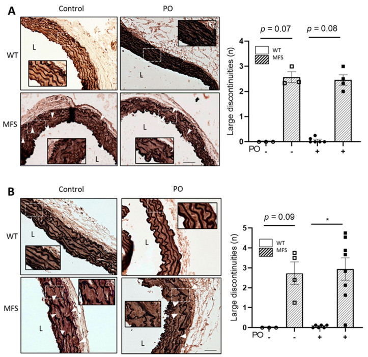
Figure 4.
Aortic wall organisation in WT and MFS mice treated with potassium oxonate. Representative light microscope images of elastin histological staining (elastin Verhoeff-Van Gieson) of aortic paraffin sections of the tunica media of the ascending aorta from WT and MFS mice treated or not with potassium oxonate (PO) after 4 weeks (A) and 16 weeks (B). The quantitative analyses of aortic elastic breaks are also shown beside their respective images. Results are the mean ± SEM. Statistical analysis: Kruskal-Wallis, Dunn’s post hoc, * p ≤ 0.05.

