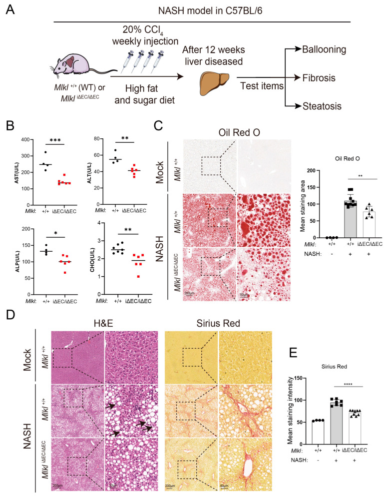
Figure 2.
Deletion of endothelial MLKL alleviates histopathological phenotype associated with NASH progression in mice. (A) Schema depicting approach to build a mouse NASH model and the indicators to describe progression of NASH. (B) Concentrations of serum AST, ALT, ALP, and CHO in Mlkl+/+ and MlkliΔEC/iΔEC NASH model. n ≥ 4 per group. AST: aspartate amino transferase, ALT: alanine aminotransferase, ALP: alkaline phosphatase, CHO: cholesterol. (C) Oil Red O staining was carried out and the result was quantified in Mlkl+/+ and MlkliΔEC/iΔEC mice with corresponding groups. Left: ROI (region of interest, 8×), scale bars: 200 μm. Right: Magnified images of indicated dotted box areas (40×), scale bars: 50 μm. (D) Liver histopathology analyzed by H&E, Sirius Red staining, and quantification (E) of Mlkl+/+ mice and MlkliΔEC/iΔEC mice with corresponding groups. Left: ROI (region of interest, 8×), scale bars: 200 μm. Right: Magnified images of indicated dotted box areas (40×), scale bars: 50 μm. Black arrow: Ballooned hepatocytes. n ≥ 4 per group. Data are shown as means ± SEMs. * p < 0.05, ** p < 0.01, *** p < 0.001, **** p < 0.0001.

