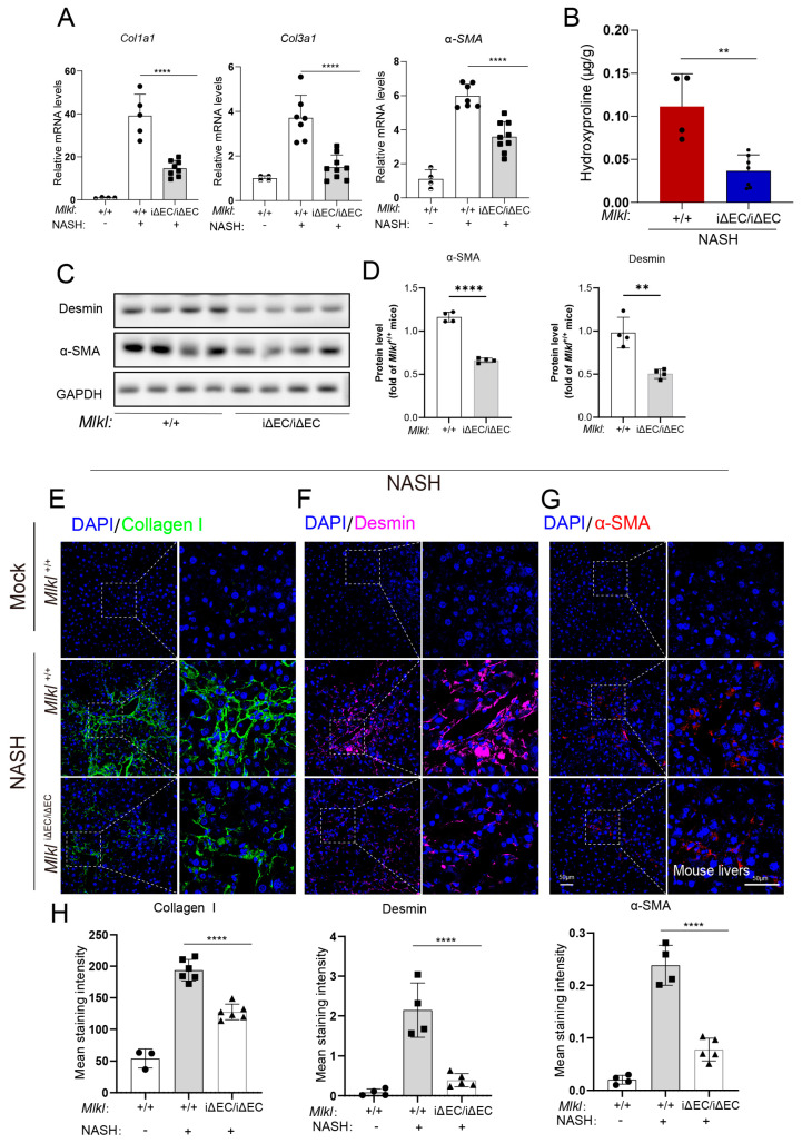
Figure 3.
Deletion of endothelial MLKL mitigates fibrosis in NASH model. (A) Relative mRNA level of Col1a1, Col3a1, and α-SMA in liver tissue were quantified by RT-qPCR showing there was a reduction in MlkliΔEC/iΔEC mice compared with Mlkl+/+ mice in NASH model. n ≥ 4 per group. (B) Hydroxyproline assay was assessed in Mlkl+/+ and MlkliΔEC/iΔEC NASH model. n ≥ 4 per group. (C) Protein levels of Desmin, α-SMA in Mlkl+/+, and MlkliΔEC/iΔEC NASH model was shown by Western blot and quantified (D). (E–G) Immunostaining for Collagen I (green), Desmin (purple), α-SMA (red), and DAPI (blue, nucleus) in Mlkl+/+ and MlkliΔEC/iΔEC NASH model. Left: ROI (region of interest), scale bars: 50 μm. Right: Magnified images of indicated dotted box areas, scale bars: 50 μm. (H) The mean staining intensity were quantified by image J (H). n ≥ 4 per group. Data are shown as means ± SEMs. ** p < 0.01, **** p < 0.0001.

