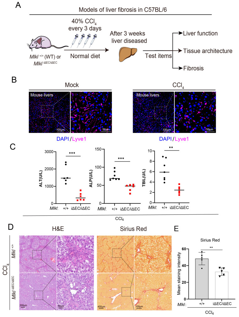
Figure 4.
Deficiency of endothelial MLKL improves the liver function and mitigates inflammation and fibrosis in CCl4 model. (A) Approach to generating a liver fibrosis model in mice. (B) Representative images of immunostaining for Lyve1 (purple) in liver sections. Left: ROI (region of interest, 20×), scale bars: 100 μm. Right: Magnified images of indicated dotted box areas, scale bars: 50 μm. (C) Serum content of liver function index including ALT, ALP, and TBIL in Mlkl+/+ and MlkliΔEC/iΔEC fibrosis model. ALT: alanine aminotransferase, ALP: alkaline phosphatase, total bilirubin. n ≥ 3 per group. (D) H&E and Sirius Red staining in Mlkl+/+ and MlkliΔEC/iΔEC mice after CCl4 injection. Left: ROI (region of interest, 8×). Right: Magnified images of indicated dotted box areas (40×), scale bars as shown in pictures. (E) Quantification of Sirius Red staining shown. n ≥ 5 per group. Data are shown as means ± SEMs. ** p < 0.01, *** p < 0.001.

