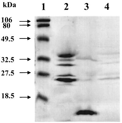
FIG. 2.
Localization of c-type cytochromes of A. faecalis. Samples were subjected to SDS-PAGE on a 15% polyacrylamide gel. An approximately equivalent amount of each cell fraction on a per-cell-weight basis was loaded onto the gel. The gel was specifically stained for heme-containing proteins as previously described (6). Lane 1, Bio-Rad prestained molecular weight markers; lane 2, membrane fraction; lane 3, periplasmic fraction; lane 4, cytoplasmic fraction.

