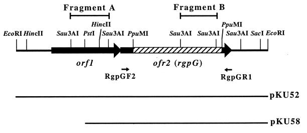
FIG. 1.
Restriction map of the EcoRI 2.6-kb insert fragment cloned in pKU52. The large arrows indicate the locations of the two ORFs. The hatched bar shows the 0.9-kb PpuMI fragment that was replaced with the Emr gene in Xc52. The small arrows labelled RgpGF2 and RgpGR1 show the positions of primers RgpGF2, 5′-GATAAGCTAGATGATACCTT-3′ (complementary to positions 1078 to 1097), and RgpGR1, 5′-AAATTACTTTTTCTTCTTAC-3′ (complementary to positions 2244 to 2225), respectively. The locations of the inserted fragments are indicated in the lower portion of the diagram.

