Figure 1.
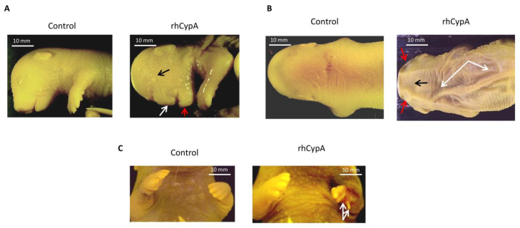
Development of anatomical anomalies in fetuses after systemic injection of rhCypA into pregnant females in the period of organogenesis. Female F1(CBA/Lac × C57BL/6) mice were mated with F1(CBA/Lac × C57BL/6) males. The next morning, females were selected by a copulative plug and s.c. injected with 2.0 mg/mice rhCypA during the period of organogenesis (6.5–11.5 dpc). Control mice received PBS for the whole gestation period. Females were sacrificed on 18.5 dpc, and the anatomical anomalies of the fetuses were visually examined. (A) Anatomical anomalies in the facial region of the embryo’s head: an underdeveloped eye (black arrow), defective maxilla and nose (white arrow), and impaired development of the mandible (red arrow). (B) Anatomical anomalies in the cranial region of the embryo’s head: deformations of the skull in the zone of the parietal bone (red arrows) and the interparietal bone (black arrow); excessive skin folds on the embryo’s back (white arrows). (C) Defects in the distal region of the forelimb: anomalies in the development of the fourth and fifth metacarpals and corresponding phalanges (white arrows).

