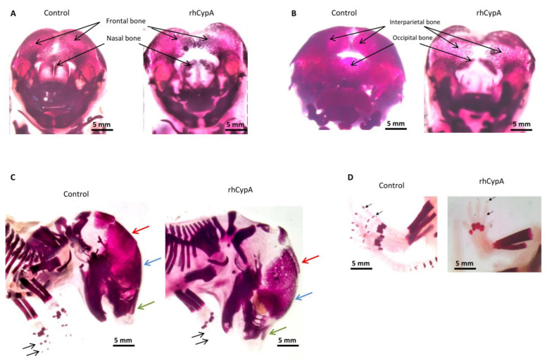
Figure 3.
Skeletal anomalies in fetuses after systemic administration of rhCypA to pregnant females in the period of organogenesis. Female F1 (CBA/Lac × C57BL/6) mice were mated with male F1(CBA/Lac × C57BL/6) mice. The next morning, females were selected by a copulative plug and s.c. injected with 2.0 mg/mice rhCypA within the period of organogenesis (6.5–11.5 dpc). Control mice received PBS for the whole gestation period. Females were sacrificed on 18.5 dpc, fetuses were stained with alizarin red, and skeletal anomalies were visually examined. (A) Impaired ossification and development of the frontal and nasal bones. (B) Anomalies in the development of the interparietal and occipital bones. (C) Defects in ossification and development of the parietal bones (red arrow). Defective ossification of the frontal (blue arrow) and nasal (green arrow) bones. Absence of distal phalanges on the forelimbs (black arrows). (D) Absence of distal phalanges on the hind limb (black arrows).

