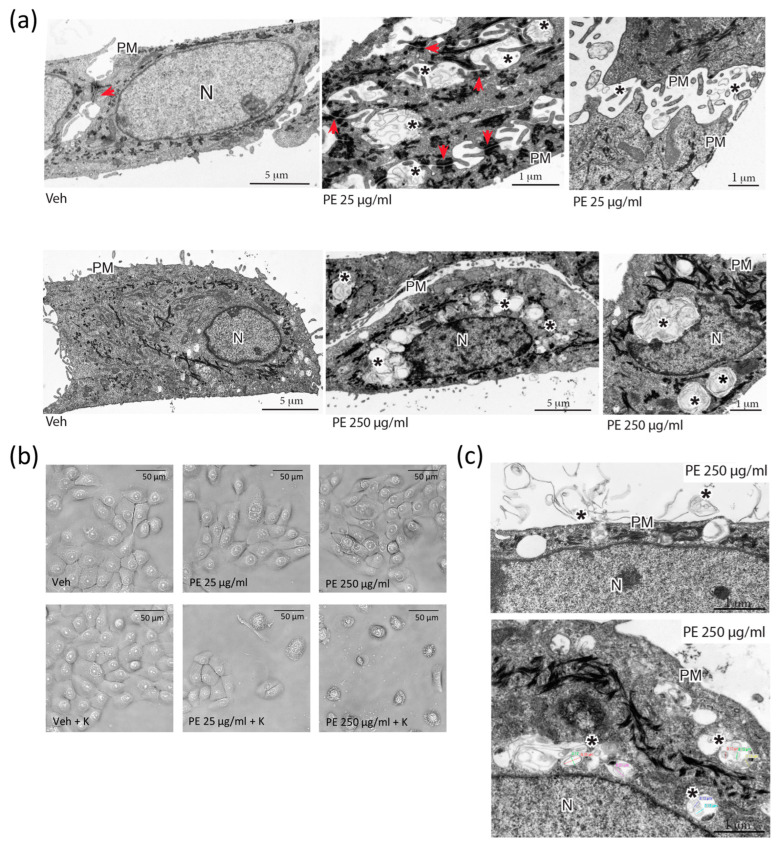
Figure 2.
Assessment of cellular uptake of PE N/MPLs by vaginal keratinocytes upon acute exposure. (a) TEM analysis of VK2 E6/E7 cells after 72 h of treatment with 25 and 250 μg/mL of PE N/MPLs or vehicle. Upper panel: From left to right, VK2 E6/E7 cells exposed to vehicle versus 25 μg/mL of PE N/MPLs. In the latter, extracellular accumulation of N/MPLs (asterisks) is evident between cells, in proximity to surface microvilli and intercellular junctions (red arrows). Lower panel: From left to right, VK2 E6/E7 cells exposed to vehicle versus 250 μg/mL of PE N/MPLs. In the latter, the N/MPLs (asterisks) are principally localised within cytoplasmic endocytic structures, often distributed around the nucleus. Two different vehicle pictures are present in the panel, since we performed the experiment with two distinct vehicle volumes matching the two PE N/MPLs concentrations. (b) Optical microscopy analysis of VK2 E6/E7 cells exposed for 72 h to vehicle versus PE N/MPLs coated with 0.2% human keratin in aqueous solution. (c) Ultrastructural analysis of the uptake of PE N/MPLs (250 μg/mL) coated with 0.2% human keratin in aqueous solution. TEM micrographs show PE N/MPLs (asterisks) deposited on the plasma membrane of VK2 E6/E7 cell, with initial uptake in forming vacuoles (top), and internalisation within peripheral and perinuclear endosome-like organelles (bottom). The NP mean diameter, reported with coloured lines in the figure (bottom), is consistent with that of the N/MPLs used in the experiment. PM: plasma membrane; N: nucleus. Scale bar: 1 μm.

