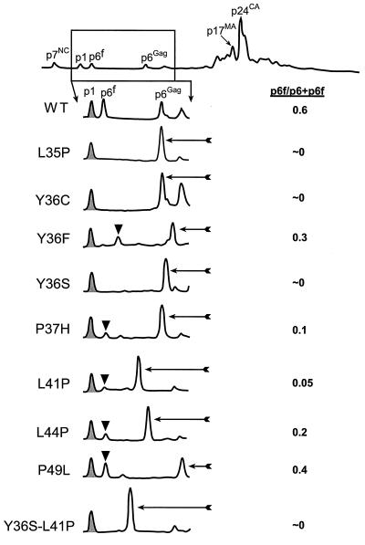
FIG. 3.
HPLC analysis of p6Gag mutants. The complete HPLC chromatogram for wild-type NL4-3 virions is shown at the top, with the region presented below boxed. The identities of the Gag proteins determined by C-PAGE, immunoblotting, protein sequence, and mass spectrometry are identified above the peaks. The p1Gag peak is shaded; the p6Gag fragment peak, labeled p6f on the wild-type (WT) profile, is identified by a black triangle; full-length mutant p6Gag is highlighted with an arrow. The relative amount of p6Gag cleaved versus the total p6Gag present (all three species) is presented on the right.

