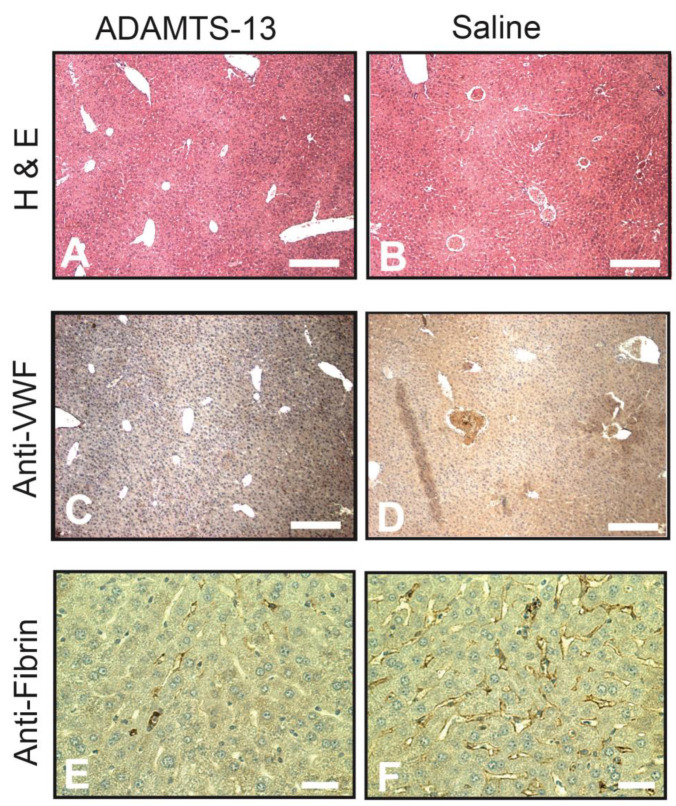
Figure 5.
Immunohistochemistry: the liver was collected from mice sacrificed 6 h after LPS injection, sectioned, and stained for H&E ((A,B), bar = 100 µm), VWF ((C,D), bar = 100 µm), and fibrin ((E,F), bar = 20 µm) on an automatic histochemistry-staining platform. Images are representative of sections from 25 mice in each experimental group.

