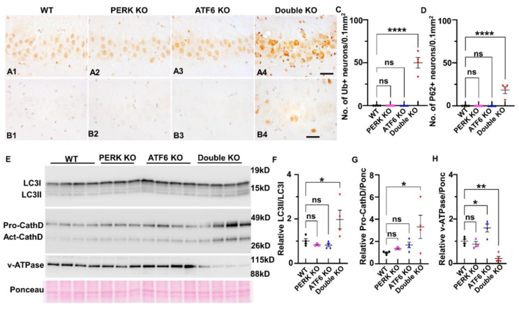
Figure 8.
Inactivation of PERK and ATF6α led to impairment of the ALP. (A1–A4) ubiquitin IHC showed an elevated level of ubiquitin in the hippocampus of Double KO mice at PID60 compared to WT mice, ATF6 KO mice, and PERK KO mice. Scale bar, 20 μm. (B1–B4) p62 IHC showed an elevated level of p62 in the hippocampus of Double KO mice at PID60, as compared to WT mice, ATF6 KO mice, and PERK KO mice. Scale bar, 20 μm. (C,D) Quantification of Ubiquitin, and P62 positive neurons in the hippocampus of WT mice, ATF6 KO mice, PERK KO mice, and Double KO mice. N = 4 mice for each group. Data are presented as means ± s.e.m. ns: no significance; **** p < 0.0001. (E,F) Western blot showed the elevated level of LC3-II/LC3-I in the forebrain of Double KO mice at PID60, compared to WT mice, ATF6 KO mice, and PERK KO mice. (E,G) Western blot showed the elevated level of pro-cathepsin D (pro-CathD) in the forebrain of Double KO mice at PID60, compared to WT mice, ATF6 KO mice, and PERK KO mice. (E,H) Western blot showed the decreased level of ATPase V0a1 in the forebrain of Double KO mice at PID60, as compared to WT mice, ATF6 KO mice, and PERK KO mice. WT, N = 4 mice; PERK KO, N = 3 mice; ATF6 KO, N = 4 mice; Double KO, N = 4 mice. Data are presented as means ± s.e.m. ns: no significance; * p < 0.05, ** p < 0.01, **** p < 0.0001.

