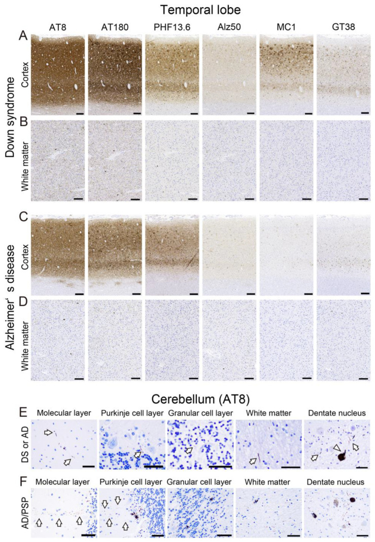
Figure 4.
Representative microphotographs of tau pathology in the temporal lobe and cerebellum of DS and sAD. (A–D) Temporal cortex; (E,F) cerebellum (AT8). (A,B) DS case (Case No. DS1; see Table 1); (C,D) sAD case (Case No. sAD5; see Table 1). (F) sAD case with progressive supranuclear palsy (PSP) pathology (AD/PSP; Case No. sAD10; See Table 1). In the temporal lobe, cortical and white matter tau deposition is generally more severe in the DS case than in the sAD case. In contrast, in the cerebellum, only small thread-like (arrows) or dot-like lesions are observed in the molecular layer, Purkinje cell layer, granular cell layer, and WM in both DS and AD cases. In the dentate nucleus, threads and neuronal intracytoplasmic tau immunoreactivity are observed (arrowhead). (F) In the patient with PSP, band-like tau immunoreactivity perpendicular to the surface is observed in the ML (arrow). In addition, astrocytic tau deposition is observed in the Purkinje cell layer. Note the dot-shaped tau deposits in the molecular layer adjacent to the Purkinje cell layer lesion (arrow). Threads and coiled bodies are identified in the granular cell layer and white matter. The tau pathology in the dentate nucleus is also more severe in this case than in the other cases. Scale bar = 250 μm (A–D), 50 μm (E,F).

