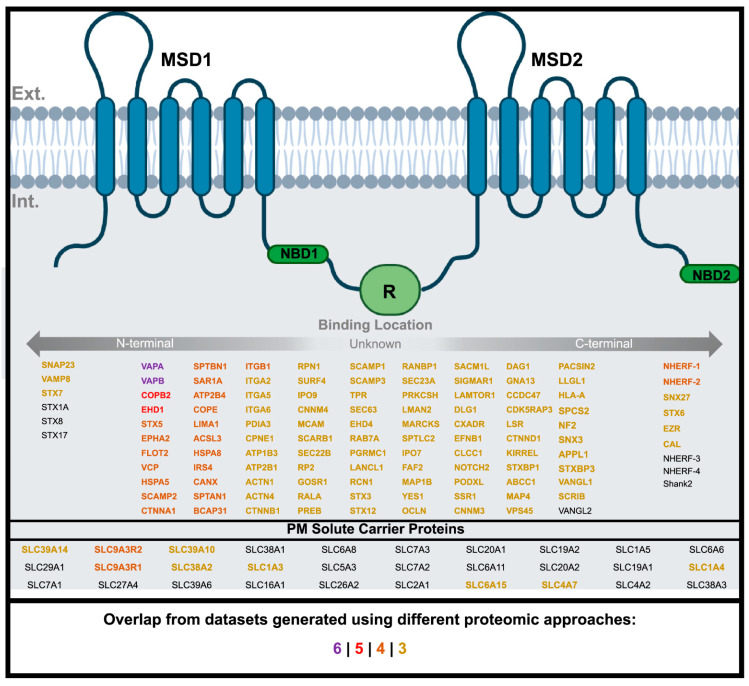
Figure 3.
WT-CFTR Surfaceome. Depiction of CFTR protein structure consisting of two nucleotide binding domains (NBD1/2), membrane spanning domains (MSD21/2), and regulatory domain (R) inserted in the PM. Interactors generated from the different proteomic approaches that are associated with the PM according to Gene Ontology (Supplementary Dataset S5), arranged according to their predicted (if known) site of binding to CFTR. Degree of overlap of interactors between different approaches is represented by colour: purple (6), red (5), orange (4), and yellow (3). Created using BioRender.com.

