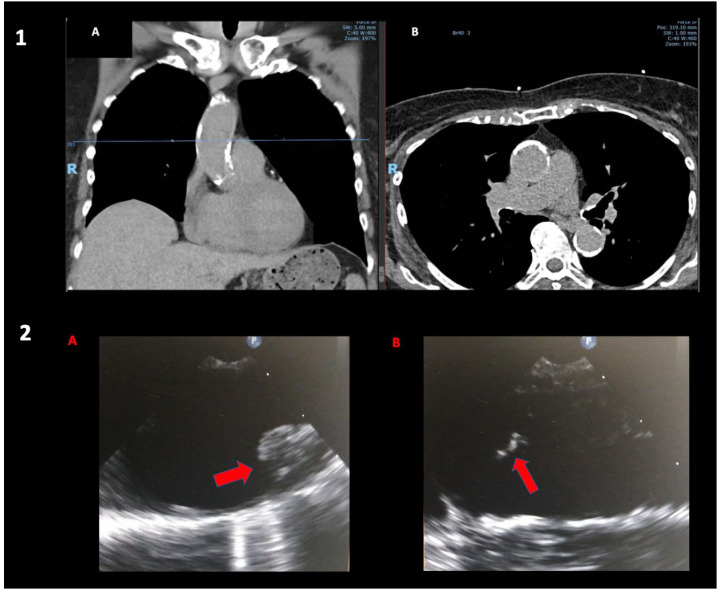
Figure 1.
(Panel 1): Coronal (A) and axial (B) views of a chest CT showing extensive ascending aortic calcifications in a 75-year-old lady admitted with unstable angina; coronary angiogram showed severe distal left main disease. (Panel 2): Intraoperative TOE showing grade IV (>5 mm) (A) and grade 5 (mobile) (B) aortic arch atheroma of a 72-year-old man undergoing combined right carotid endarterectomy and anaortic OPCAB.

