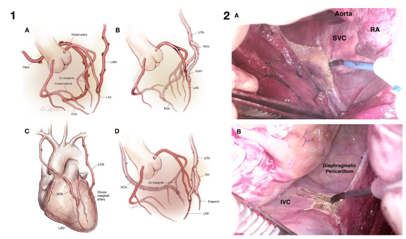
Figure 3.
(Panel 1): Common configurations of composite arterial grafts. (A) LITA to LAD and RITA-LRA extension though the transverse sinus to the lateral and inferior system [1]; (B) LITA to LAD and RITA as a Y graft from LITA to the lateral and inferior systems [33]; (C) in situ RTA to LAD and LITA to obtuse marginal [34]; (D) LITA to LAD, LRA as a Y graft from LITA to diagonal branch and RITA to obtuse marginal [33]. Figures reprinted according to CC BY-NC-ND 4.0 license. (Panel 2): (A) Right superior pericardial slit. During a brief period of apnea, a vertical pericardial slit is made with diathermy down to and including the pericardial fold at the right atrial/SVC junction. The assistant retracts the thymus with their right hand using the Yankeur sucker head and retracts the aorta using reversed De Bakey forceps in their left hand. Extreme care must be taken not to injure the right phrenic nerve. (B) Right inferior pericardial slit. A vertical pericardial incision is made with diathermy down to the IVC. Care is taken to remain extra-pleural and to avoid injury to the right phrenic nerve [35]. Figures reprinted according to CC BY-NC-ND 4.0 license. (LITA: left internal thoracic artery; RITA: right internal thoracic artery; LRA: left radial artery).

