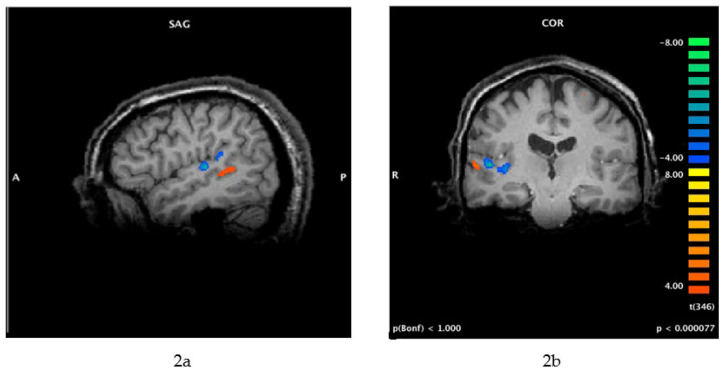
Figure 2.
(a,b) Functional MRI (fMRI) of brain activity associated with tinnitus (this patient perceived 12,000 Hz tinnitus on the right side only). Cortical activity associated with tinnitus perception (orange) is more lateral (within secondary auditory cortex) compared with the neural activity in the primary auditory cortex that is associated with the perception of external sounds (blue). These images were obtained using a protocol that included residual inhibition, as follows: The scan began with a 30 s resting baseline, followed by 1 min of masking noise. The patient pressed one button when he perceived that his tinnitus was effectively masked, another button when his tinnitus began to return after masking, and a third button when his tinnitus was back to its original loudness. SAG = sagittal view; COR = coronal view; A = anterior; P = posterior; R = right.

