Figure 2.
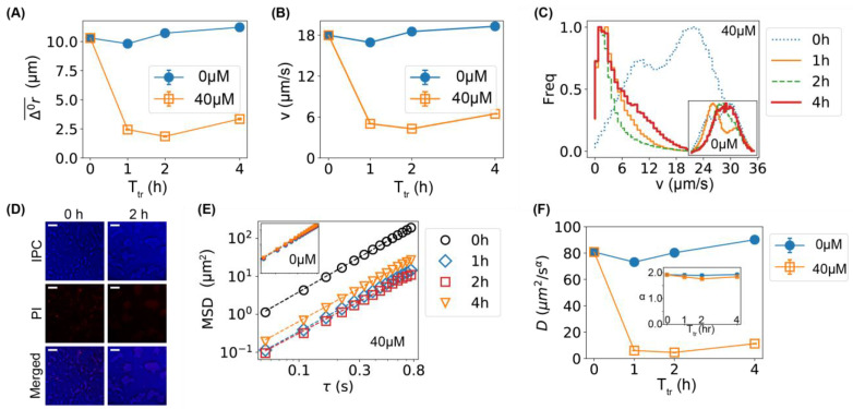
Lower motility of bacteria caused by Ag+ ions. (A) The dependence of the mean displacements () of the first 12 frames of all trajectories of bacteria on incubation/treatment time in the absence (0 µM) and presence of Ag+ ions (40 µM). (B) The dependence of the mean bacterial velocity on incubation/treatment time in the absence (0 µM) and presence of Ag+ ions (40 µM). (C) Distributions of bacterial velocities in the presence of Ag+ ions at 40 µM for 0, 1, 2, and 4 h. Inset: the corresponding result for untreated bacteria (0 µM). The axes of the inset are the same as the main figure. (D) Cell-viability assay based on propidium iodide (PI) staining for untreated (0 h, left column) and treated (2 h, right column) bacteria. Top: inverted phase-contrast (IPC) images; middle: fluorescence images due to PI staining; bottom: merged IPC/PI images. Scale bar = 16 µm. (E) Log–log plot of mean-square displacements (MSDs) vs. lag time () for trajectories of treated bacteria by Ag+ ions at 40 µM for 0, 1, 2, and 4 h. Inset: the corresponding result for untreated bacteria (0 µM). (F) Dependencies of the generalized diffusion coefficient D and the anomalous scaling exponent (inset) on the incubation/treatment time Ttr.

