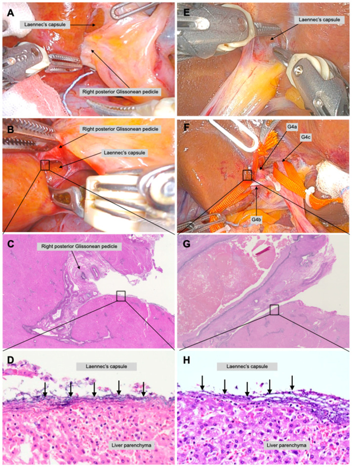
Figure 3.
Robotic second and third branches of Glissonean pedicle isolation in a live body. Right posterior Glissonean pedicle isolation was performed during robotic right posterior sectionectomy. The junction of the right anterior and posterior Glissonean pedicles was identified and exposed the Laennec’s capsule (A). Blunt dissection between the Laennec’s capsule and the right posterior Glissonean pedicle from the caudal view while directly observing the dorsal view with a robotic endoscope (B). The Laennec’s capsule on the liver parenchyma and the exfoliated right posterior Glissonean pedicle of a live body is shown in low- (C) and high-power fields (black arrows) (D). Glissonean pedicle 4a (G4a), 4b (G4b), and 4c (G4c) isolation was performed in robotic segmentectomy 4b. The gap between the umbilical plate and the Laennec’s capsule was separated (E). All responsible branches of the Glissonean pedicle can be isolated without misidentification while directly observing the branches (F). The Laennec’s capsule on the liver parenchyma and the exfoliated Glissonean pedicle is in low- (G) and high-power fields (black arrows0 (H).

