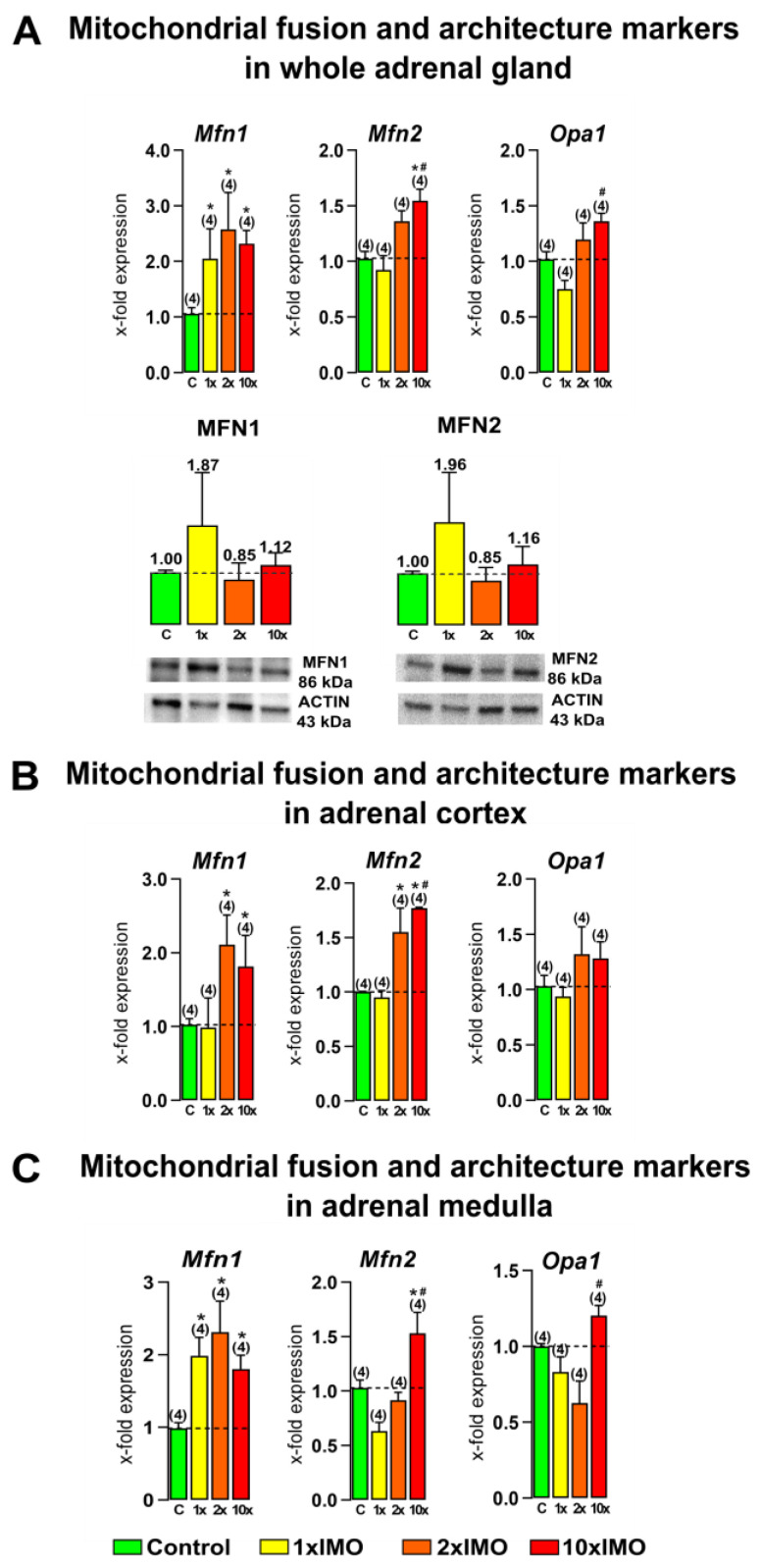
Figure 3.
Ten repeated psychophysical stress incidents increased the expression of mitochondrial fusion and architecture markers in the whole adrenal gland, adrenal cortex, and medulla tissues. Whole adrenal gland tissue (A), as well as the adrenal cortex (B) and medulla (C) tissues, were isolated from undisturbed rats, as well as acutely (1 × IMO) and repeatedly (2 × IMO and 10 × IMO) stressed rats. Tissue was further used for RNA isolation followed by the analysis of the transcriptional profile of mitochondrial fusion markers. Whole adrenal gland tissue was used for MFN1 and MFN2 protein expression analyses. The representative blots are shown as panels. Data from scanning densitometry were normalized to ACTIN (endogenous control). Values are shown as bars above the photos of blots. The data bars represent mean ± SEM values of two independent in vivo experiments (number in brackets above the bars represents number of analyzed animals). Statistical significance was set at level of p < 0.05: * vs. control group and # vs. 1 × IMO group.

