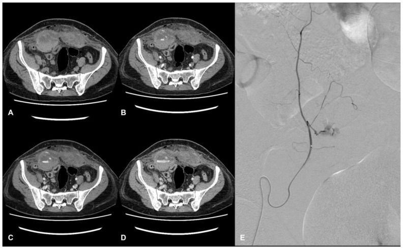
Figure 1.
CT and DSA images of a hospitalized patient with COVID-19. Evidence of a large hematoma within rectus abdominis below the arcuate line in axial CT images. The pre-contrast phase shows the extension and location of the hematoma, showing a fluid level (hematocrit effect) (A). The contrast-enhanced acquisition at the arterial (B), portal (C), and delayed phase (D) show active bleeding within the hematoma. Angiography shows a contrast blush from branches of the right inferior epigastric artery (E).

