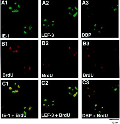
FIG. 4.
Double staining of BmNPV-infected BmN cells with BrdU and IE-1, LEF-3, or DBP. (A) Immunofluorescence images of BmNPV-infected BmN cells at 8 h p.i. with antibodies against IE-1 (A1), LEF-3 (A2), or DBP (A3). For IE-1 staining, FITC-conjugated goat anti-guinea pig IgG was used. For DBP and LEF-3 staining, FITC-conjugated goat anti-rabbit IgG was used. (B) BrdU immunofluorescence images of the same cells as in panel A. For BrdU staining, rhodamine red X-conjugated goat anti-mouse IgG was used. (C) Panels A and B merged.

