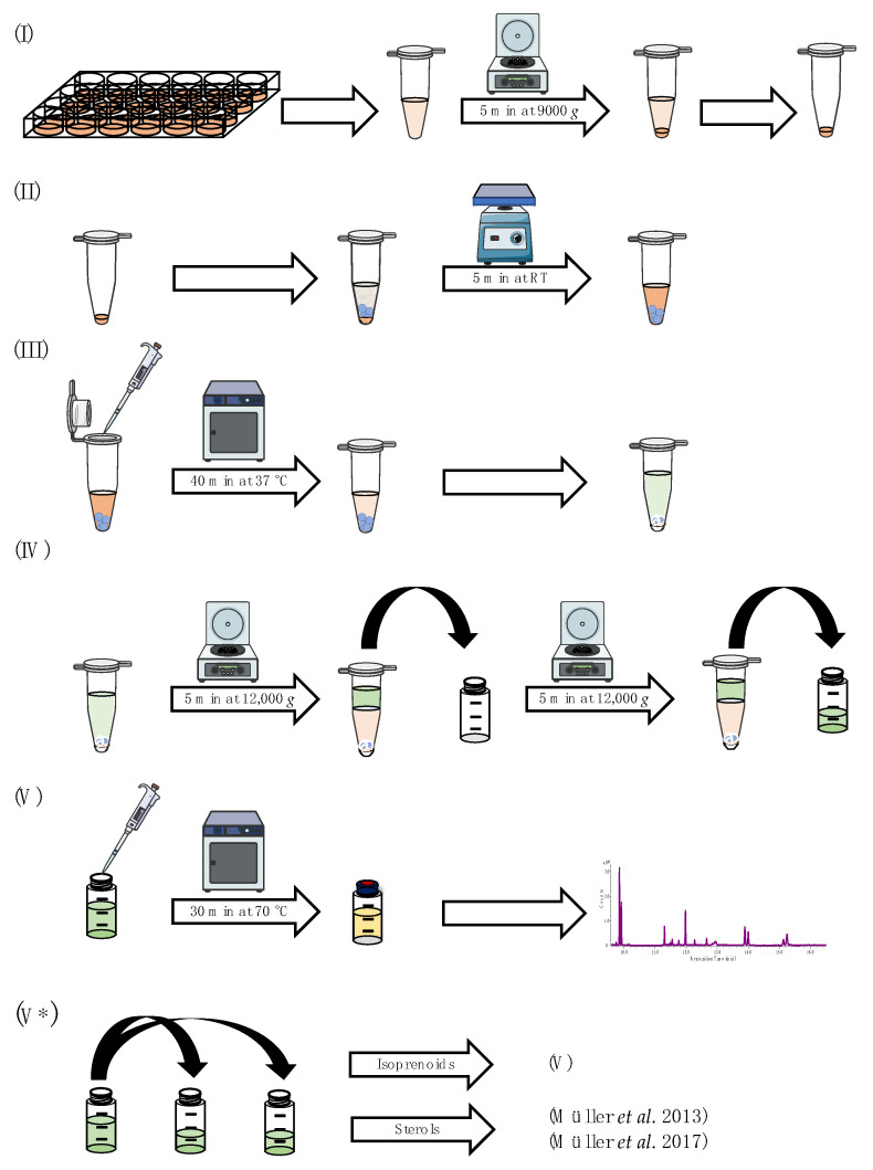
Figure 2.
Schematic overview of the sample preparation, which can be divided into five individual steps: (I) preparation of cellular matrix; (II) mechanical lysis of cells/lyophilized mycelia in enzymatic buffer; (III) enzymatic deconjugation and preparation of liquid–liquid extraction; (IV) liquid–liquid extraction; (V) derivatization and GC-MS measurement; (V*) alternative sample preparation for additional sterol pattern analysis [9,28]. Parts of this figure were created using Servier Medical Art templates, licensed under a Creative Commons Attribution 3.0 Unported License (https://smart.servier.com (accessed on 19 July 2023).

