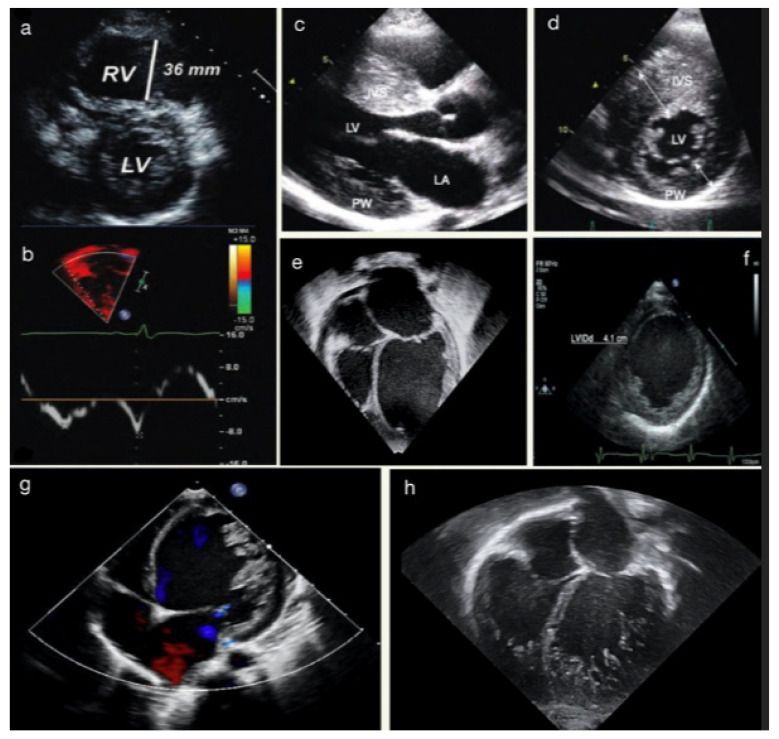
Figure 1.
(a) Parasternal short-axis view in a 14-year-old female with ventricular tachycardia during exercise, demonstrating right ventricular (RV) enlargement with suspected ACM. (b) TDI of the RV free-wall showing diminished tricuspid annular TD velocities of the same patient. (c) Parasternal long-axis view in a 13-year-old male with SIV hypertrophy in HCM. (d) Parasternal short-axis view showing hypertrophy of mid-ventricular IVS. (e) Apical-4-chamber view showing dilated LV and LA in suspected DCM. (f) Parasternal short-axis view demonstrates LVEDD at the papillary muscles level. The images (g,h) show two different cases of LVNC.

