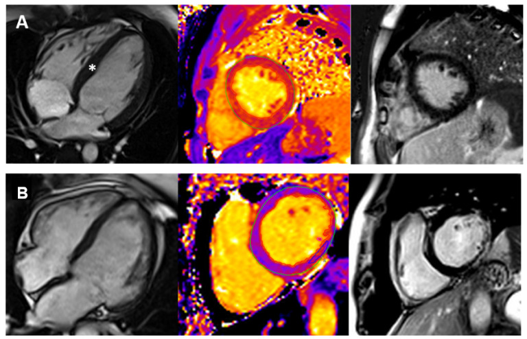
Figure 2.
Cardiovascular Magnetic Resonance Imaging in three patients with different cardiomyopathy phenotypes. Panel (A): early hypertrophic cardiomyopathy (HCM). There is concentric left ventricular hypertrophy (left), accompanied by prolonged T1 mapping values (center) and no late gadolinium enhancement. The white asterisk (left) indicates the hypertrophic septum. Panel (B): dilated cardiomyopathy (DCM) in a patient with Duchenne muscular dystrophy. There is moderate chamber enlargement and LV systolic dysfunction (LVEF 37%) (left). T1 mapping values are globally elevated (center), and there is mainly epicardial late enhancement of the basal to mid lateral wall (right).

