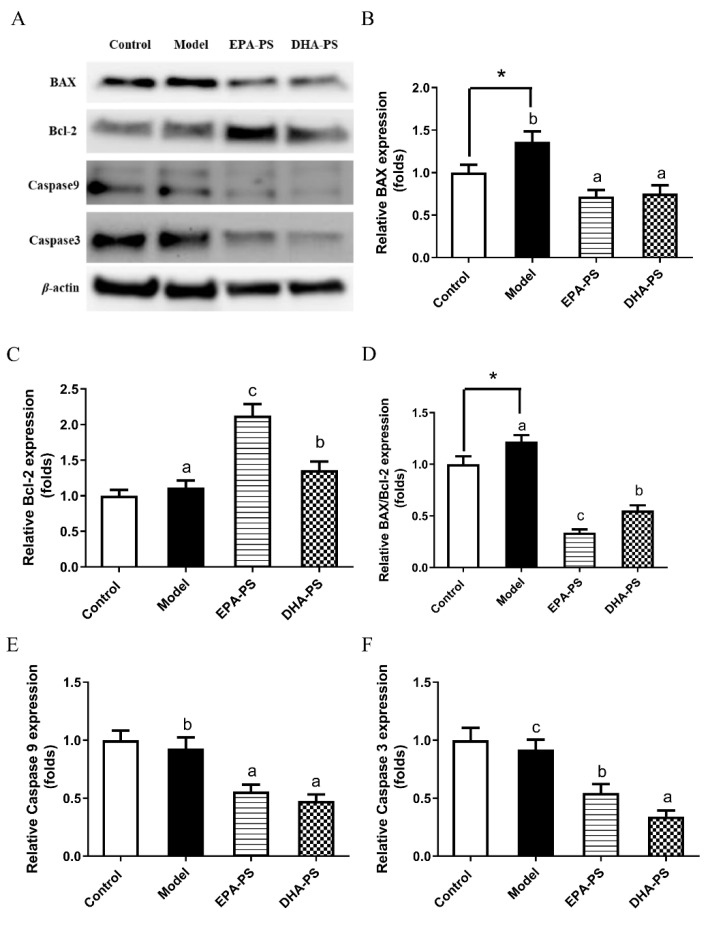
Figure 3.
Effects of EPA-PS and DHA-PS on the expression of apoptotic proteins in primary hippocampal neurons after oxidative damage. Representative western blots (A) and relative expression of BAX (B), Blc-2 (C), BAX/Blc-2 (D), Caspase 9 (E) and Caspase 3 (F) in primary hippocampal neurons. The expressions were detected by Western blotting analysis and normalized with β-actin. * p < 0.05 indicates significant differences compared with the control group. Different letters represent significant differences at p < 0.05 among treated groups. This figure shows the mean ± SEM of 3 experiments.

