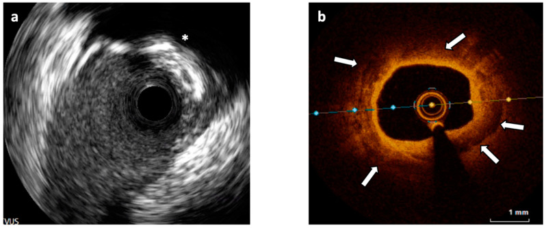
Figure 2.
(a) Intravascular Ultrasound (IVUS) cross-section showing a 180° calcified plaque opposite to a side branch origin and producing shadow (asterisk). (b) Optical Coherence Tomography (OCT) cross-section of a different segment of the vessel showing an almost concentric calcified plaque visible as signal-poor regions with sharply delineated borders (white arrows).

