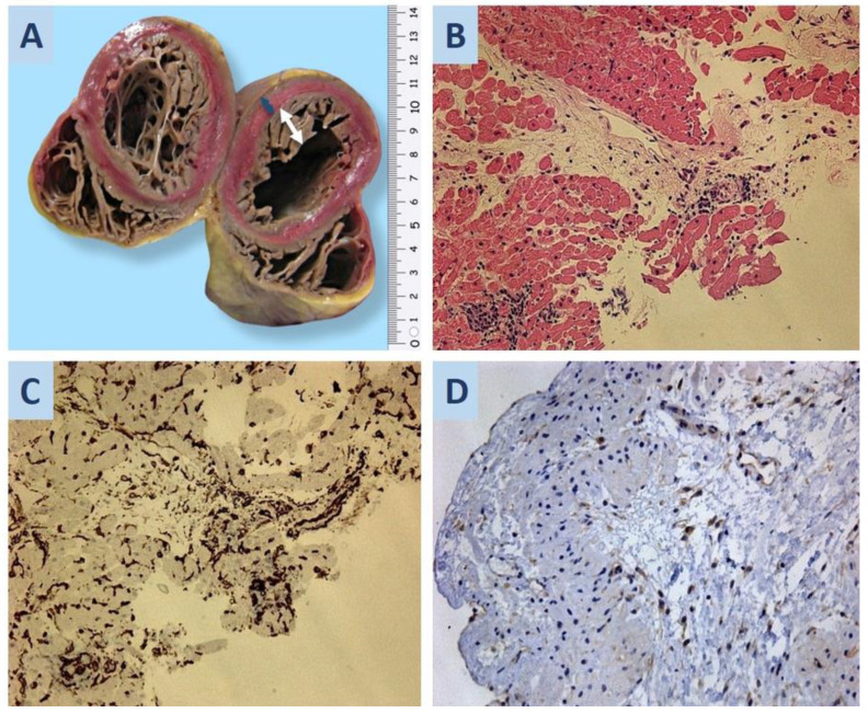
Figure 2.
(A) Noncompact myocardium of the left ventricle: the blue arrow shows the thickness of the compact layer (0.6 cm), and the white arrow shows the non-compact layer (1.6 cm). (B) Second EMB of the donor heart: a visible infiltrative vasculitis with macrophages in the lumen of blood vessels; hematoxylin-eosin staining; ×200. (C) Immunohistochemical staining shows CD34 expression in large, polymorphic endothelial cells; ×200. (D) SARS-CoV-2 S-protein expression in the cytoplasm of large, polymorphic endothelial cells; ×200.

