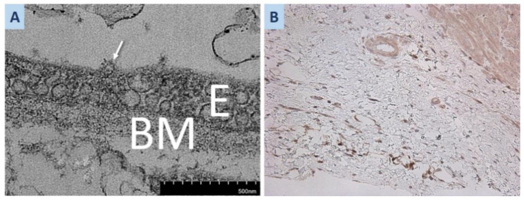
Figure 4.
(A) Second EMB of the donor heart. Round 100 nm particles with clear signs of morphological similarity to the SARS-CoV2 virion (arrow), localized at the apical surface of the endothelium. (B) SARS-CoV-2 S-protein expression in the cytoplasm of endothelial cells of the polypoid fibrolipoma myocardial site; ×100. Abbreviations: E—endothelium, BM—basal membrane.

