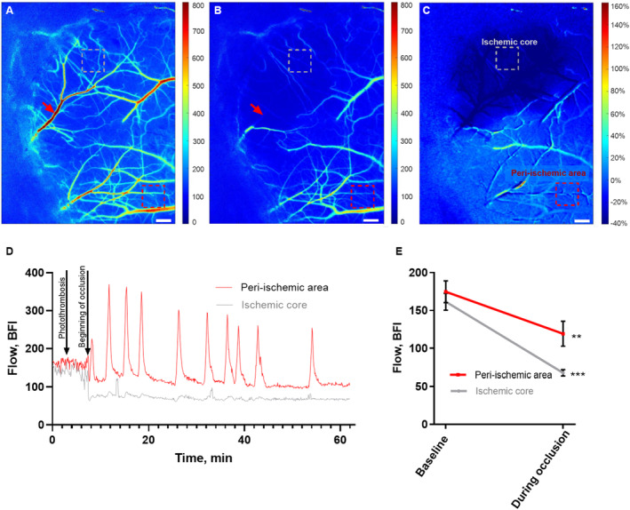
Figure 1. Cerebral ischemia induced by single‐vessel photothrombosis of the middle cerebral artery.

Representative laser speckle contrast images (LSCI) of the affected hemisphere in the awake mouse at baseline (A) and during occlusion of the anterior middle cerebral artery (MCA) branch (B). Red arrows show the focus point for the green laser beam (Ø=6 μm) used for photothrombosis of the anterior MCA branch (A and B). C, Image of the ratio of LSCI before vs during the occlusion. Bars, 300 μm; dotted boxes indicate the region of interest in the ischemic core (gray) and the peri‐ischemic area (red; A–C). D, Representative traces from these 2 regions show the changes in blood flow during the MCA occlusion. E, MCA occlusion was associated with a drop in blood flow in the ischemic core and in the peri‐ischemic area. Blood flow during occlusion was compared with baseline in 2 separate 2‐tailed paired t tests for each of the areas. Error bars=SE. **P<0.01, ***P<0.001; n=6. BFI indicates blood flow index.
