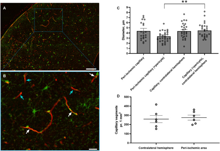
Figure 7. Diameter of capillary segments surrounded by pericyte bodies was reduced in the peri‐ischemic cortex.

Representative images from the peri‐ischemic cortex showing platelet‐derived growth factor receptor beta‐positive cells in green and lectin in red in low (A) and high magnification (B). White and turquoise arrows indicate capillary segments with and without pericyte bodies, respectively (B). Bars, 100 μm (A) and 25 μm (B). C, Measurements of capillary diameters in each hemisphere for each mouse were averaged and plotted as a bar graph. The diameter of capillaries associated with pericyte bodies in the peri‐ischemic cortex was reduced compared with the contralateral cortex. There was no difference in the diameter of capillaries that were not associated with pericyte bodies between the hemispheres. D, The density of capillary segments was similar in both hemispheres. Error bars=SE. Capillary diameter and density in peri‐ischemic cortex vs contralateral hemisphere were compared using unpaired t test. **P<0.01, n=6. See splitting of the channels including nucleus staining with 4′, 6‐diamidino‐2‐phenylindole in Figure S4.
