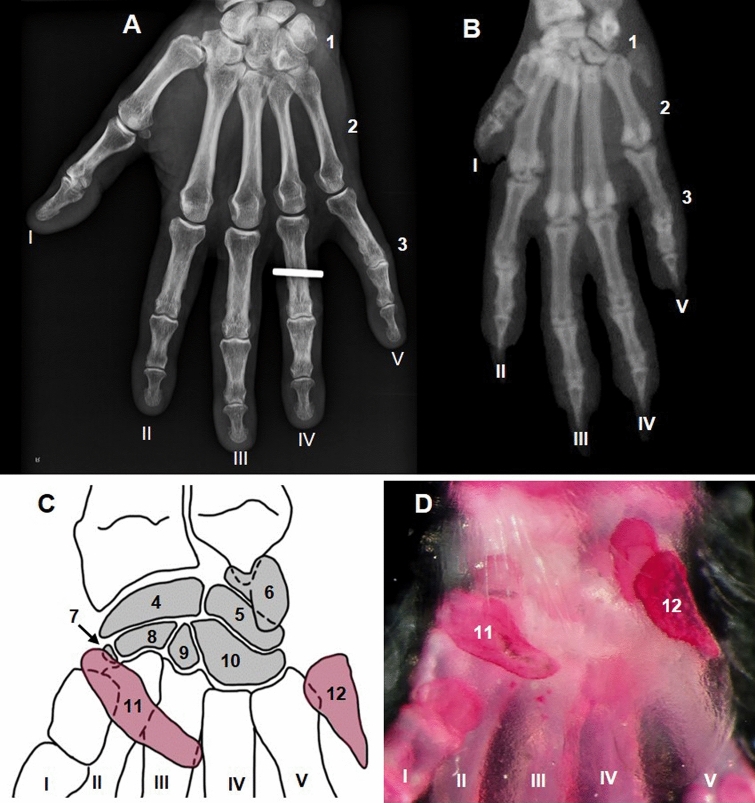
Fig. 8.
Dorsal ventral radiographs of human hand (A) and mouse forepaw (B). Diagram showing the topography and organization of mouse carpal bones (C). Alizarin stained mouse carpus (palmar aspect) (D). Carpal bones (1); metacarpal bones (2); phalanges (3); scapholunate (4); triquetral (5); pisiform (6); trapezium (7); trapezoid (8); capitate (9); hamate (10); falciform carpal bone (11), which is a sesamoid bone embedded in the flexor retinaculum and not a true carpal bone; ulnar sesamoid bone (12). Roman numerals indicate the medial to lateral order of digits

