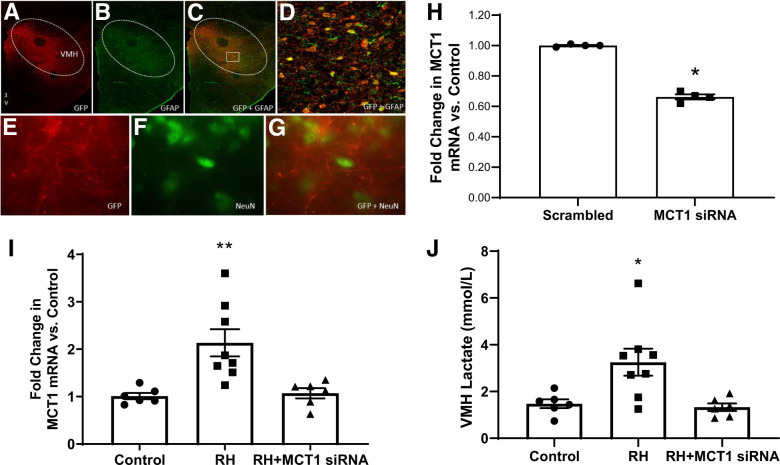
Figure 1.
(A–G) Immunohistochemical staining demonstrating cellular specificity of the AAV vector for targeting astrocytes in VMH sections. A and E show labeling of the AAV’s GFP reporter gene in red. B and F show the labeling of GFAP, an astrocyte marker or NeuN, a neuronal marker, in green. C and G are the merged images showing colocalization of the GFP signal with GFAP on top and distinct labeling of the GFP reporter and neurons (NeuN) on the bottom, indicating the AAV vector specifically targets expression to astrocytes and not neurons. D is a magnified image of the area outlined in C that more clearly shows the colocalization. (H) Quantitative RT-PCR analysis showing the MCT1 siRNA-expressing AAV (MCT1 siRNA; n = 4) decreased the expression of MCT1 mRNA in the VMH by ∼35% (*P < 0.05) compared with controls receiving the AAV expressing the scrambled RNA (Scrambled; n = 4). (I) Quantitative RT-PCR showing an increase in MCT1 mRNA expression in RH rats (squares; N = 8) compared with controls (circles; N = 6) and normalization of MCT1 mRNA levels in the VMH of RH animals using the astrocyte targeting AAV (RH+MCT1 siRNA; triangles; N = 6). (J) Extracellular lactate concentrations in the VMH were elevated in RH animals (squares), and these levels were restored to normal following knockdown of MCT1 (RH + MCT1 siRNA; triangles). RH increased extracellular lactate concentrations (squares) compared with hypoglycemia-naïve control animals (circles). Reducing astrocytic MCT1 expression in the VMH of recurrently hypoglycemic rats (triangles) decreased extracellular lactate levels to normal. Data are presented as mean ± SEM. *P < 0.05, **P < 0.01 vs. Scrambled/Control and RH+MCT1 siRNA; 3V, 3rd ventricle.

