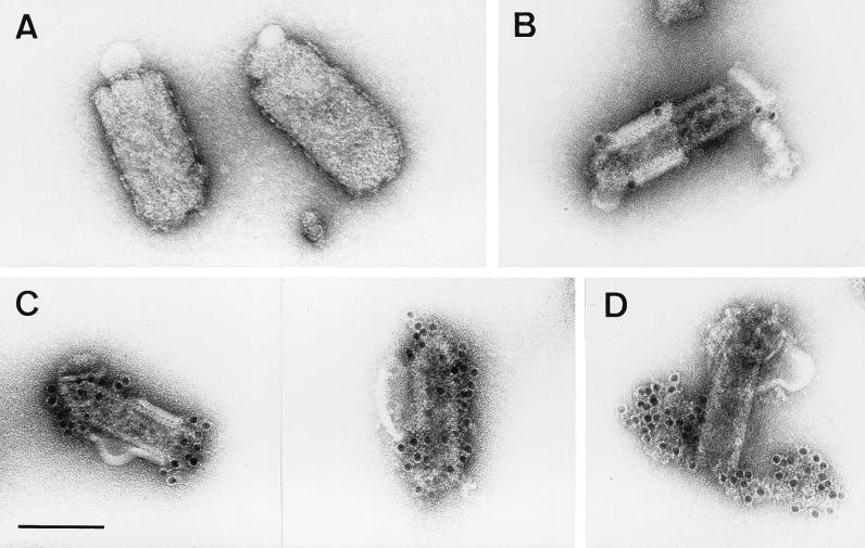
FIG. 1.
RV M protein is positioned between the lipid bilayer and the RNP coil. Virions treated with Triton X-100 (B to D) and untreated control samples (A) were purified as described in Materials and Methods and analyzed by electron microscopy. The absence of anti-M labeling on the outer membranes of untreated virions (90 to 100 nm in diameter) (A) and an intense anti-M labeling on the surfaces of the RNP coil or skeletons (50 to 60 nm in diameter) of detergent-treated virions (C) demonstrate that M surrounds the RNP coil. Only areas where the lipid bilayer was not completely removed (60 to 70 nm in diameter) are weakly labeled with anti-G MAb (B). In partially disassembled skeletons (D), only the uncoiled nucleocapsid, and not the surface of the skeleton, is labeled with anti-RNP serum. Bar, 100 nm.

