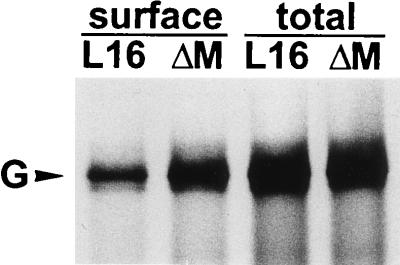
FIG. 4.
Analysis of cell surface expression of G proteins. Approximately 106 BSR cells were infected at an MOI of 1 and after 16 h were labeled with 100 μCi of [35S]methionine for 3 h. Anti-G MAb was incubated with live cells to bind G proteins expressed at the cell surface or with cell lysates containing total G protein. Phoshorimager quantitation revealed a 2.6-fold-higher level of G protein on the surfaces of cells infected by SAD ΔM.

