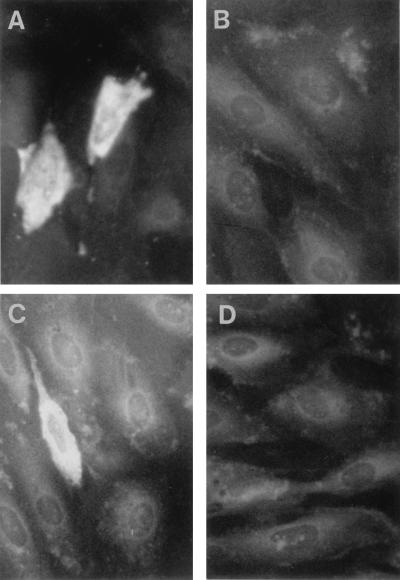
FIG. 2.
Expression of WHsAg and WHcAg in woodchuck liver cells (WH12/6) after transfection of pWHsIm and pWHcIm, respectively. WH12/6 cells were transfected with 4 μg of plasmids. After 48 h, transfected cells were fixed with acetone-methanol (1:1). The expressed WHcAg and WHsAg were detected by immunofluorescence staining with rabbit antisera to respective WHV antigens. (A) Transfection with pWHcIm and staining with anti-WHcAg antibody. (B) Transfection with pcDNA3 and staining with anti-WHcAg antibody. (C) Transfection with pWHsIm and staining with anti-WHsAg antibody. (D) Transfection with pcDNA3 and staining with anti-WHsAg antibody.

