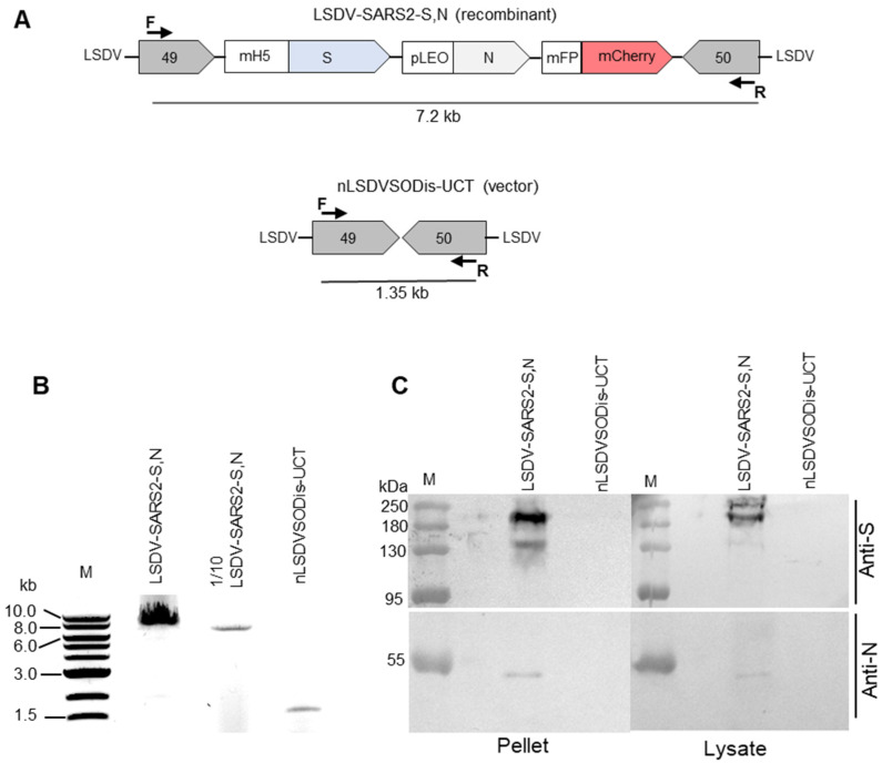
Figure 2.
Characterization of LSDV-SARS2-S,N. (A) Schematic diagram showing the PCR product sizes expected to be amplified by the forward (F) and reverse (R) primers from LSDV-SARS2-S,N and nLSDVSODis-UCT. (B) Gel electrophoresis of PCR products from samples extracted from MDBK cells infected with LSDV-SARS2-S,N or nLSDVSODis-UCT. One-tenth of the sample from the first lane was loaded in the second lane. (C) SDS PAGE and Western blot of samples from infected MDBK cells. The membrane was cut in half and probed with either anti-spike (anti-S) or anti-nucleocapsid (anti-N) antibody.

