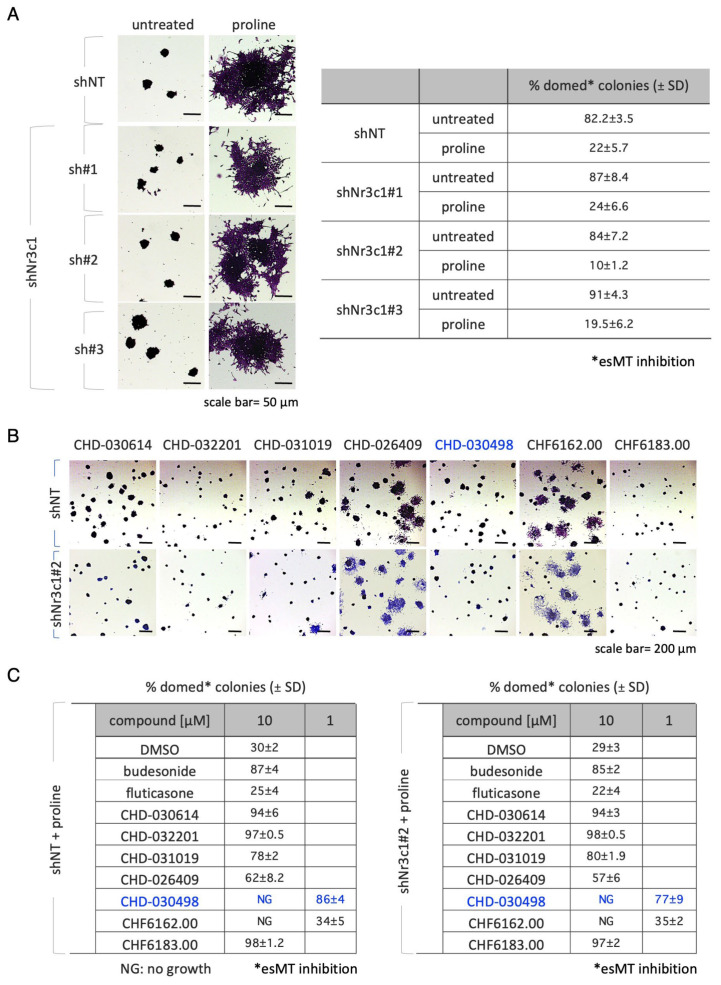
Figure 3.
Effect of glucocorticoid receptor on BA-GCs-mediated modulation of esMT. (A) Representative photomicrographs (left) of cell colonies generated from shNT (non-targeting control) and shNr3c1#2 (GR knock-down) ESCs under esMT-inducing condition (supplemental proline, 500 μM). Table showing the fraction (%) of domed-shaped cell colonies (esMT inhibition) (right). Scale bar, 50 μm. (B) Representative photomicrographs of colonies generated from shNT and shNr3c1#2 ESCs treated ± proline (500 μM) ± BA-GCs (10 μM), and stained with crystal violet. CHD-030498 was used at 1 μM. (C) Tables showing the fraction (%) of domed colonies (esMT inhibition), in the different conditions (shNT, bottom left; shNr3c1#2, bottom right). Budesonide and fluticasone were used as positive (esMT inhibitor) and negative (inactive) controls. The different effects of CHD-030498 at 10 (toxic) and 1 μM are highlighted in blue. Data are shown as mean ± SD (n = 3). Scale bar, 200 μm.

