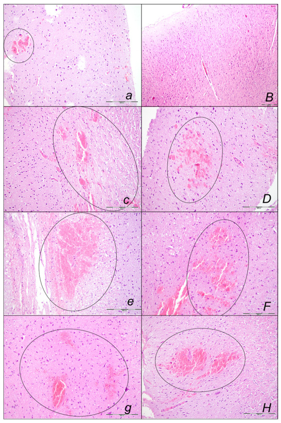
Figure 14.

Illustrative the brain hemorrhage microscopic presentation (HE staining; magnification 100×; scale bar 200 μm) in the sotalol, control rats (italic small letters) and BPC 157-treated rats (italic capitals), assessed at 15 min (a,B), 90 min (c,D) and 180 min (e,F,g,H) following application of sotalol. Medication was given as an early application immediately after sotalol (a,B,c,D,e,F), or as delayed post-treatment (application at 90 min and assessment at 180 min sotalol-time) (g,H), saline (a,c,e,g) or BPC 157 therapy (B,D,F,H) given intragastrically. Sotalol-rats treated with saline consistently had pronounced edema, congestion, and areas of intracerebral hemorrhage (a,c,e,g) with findings of multifocal hemorrhagic areas (g) (marked area) in the brain tissue affecting the deeper cortical area and white matter of the brain (e). Sotalol-rats treated with BPC 157 had mild edema and congestion of brain tissue. No hemorrhage was noted at 15 min sotalol-time (B) (period relative to therapy application 0–15 min). A lesser area of intracerebral hemorrhagic appeared at 90 min and 180 min sotalol-time affecting superficial, cortical brain area (D,F,H) (periods relative to therapy application 0–90 min, 0–180 min, 90–180 min).
