Figure 2.
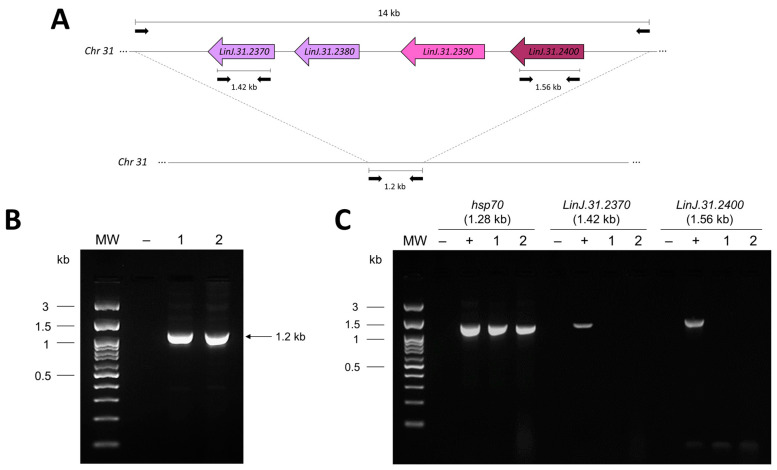
Investigation of the MSL in the L. (L.) infantum LD strain and ME clinical isolate, using PCR protocols previously described by Carnielli et al. [11]. (A) Schematic representation of the MSL of approximately 14 kb. In the absence of the MSL, a 1.2 kb DNA fragment is amplified. (B) PCR for the presence (~14 kb) or absence (~1.2 kb) of the MSL in the LD strain (lane 1) and the ME isolate (lane 2). (C) PCR amplification of the MSL genes LinJ.31.2370 (that encodes NUC1) and LinJ.31.2400 (that encodes 3,2-trans-enoyl-CoA isomerase). The size of amplified PCR products is indicated above the figure. As a control, all genomic DNAs were also evaluated by a PCR protocol that amplifies a 1.28 kb product of the hsp70 gene, as previously described by Montalvo et al. [23]. MW—molecular weight in kilobase (kb); (−) negative control for PCR (absence of genomic DNA); (+) positive control for PCR (genomic DNA of L. (L.) donovani DD8 strain); (1) L. (L.) infantum LD strain; (2) L. (L.) infantum ME clinical isolate.

