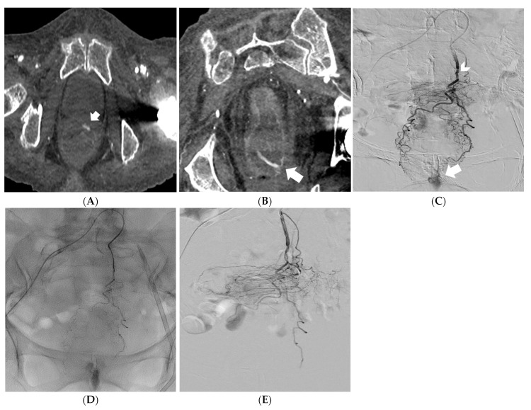
Figure 2.
Reformatted CT angiography images in the oblique axial and coronal planes showing a massive rectal hemorrhage (arrows) (A,B). Digital subtraction angiography identifying the superior rectal artery as the feeding vessel (arrowhead); active bleeding is also noted (arrow) (C). Embolization of the superior rectal artery with PVA particles mixed with iodinated contrast media under fluoroscopy control (D). Digital subtraction angiography confirming successful embolization of the feeding vessel (patency of the middle and inferior rectal arteries, fed by the internal iliac arteries, prevented significant ischemic injury of the rectal wall) (E).

