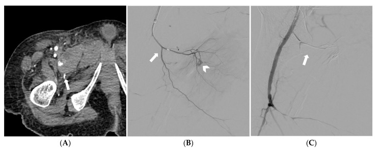
Figure 4.
CT angiography depicting a spontaneous pectineus muscle hematoma with active bleeding (arrow) (A). Selective catheterization of the right profunda femoris artery and superselective catheterization of the medial circumflex femoral artery (arrow); at digital subtraction angiography active bleeding is noted (arrowhead) (B). Digital subtraction angiography demonstrating successful embolization with a gelatin sponge (arrow); preserved patency of the profunda femoris artery and its other branches is also noted (C).

