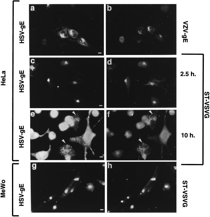
FIG. 3.
Intracellular distribution of HSV-gE in HSV-2-infected cells. (a to f) HeLa cells grown on coverslips were transfected with plasmids encoding VZV-gE (a and b) or ST-VSVG (c to f) and, 40 h after transfection, exposed to a tissue culture supernatant of MRC-5 cells infected with HSV-2 for 2 h, and then the inoculum was removed and further incubated for another 2.5 h (a to d) or 10 h (e and f). Cells were fixed and processed for indirect immunofluorescence by using the 7520 anti-HSV-gE MAb (a, c, and e) and either the 1667 polyclonal antiserum against VZV-gE (b) or a polyclonal antiserum against the VSVG epitope (d and f). (g and h) A clone of MeWo cells stably expressing ST-VSVG was exposed to a tissue culture supernatant of MRC-5 cells infected with HSV-2 for 5 h, fixed, and stained with the 7520 anti-HSV-gE MAb (g) and a polyclonal antiserum against the VSVG epitope (h). Bar, 5 μm.

