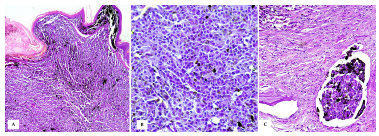
Figure 4.
Cutaneous Malignant Melanoma (A) A densely cellular neoplasm, markedly expands the dermis and reaches the dermo–epidermal junction, covered by intact or often ulcerated epidermis (HE, 20×); (B) At higher magnifications, the neoplastic cells show round to oval nuclei of varying size, moderate amount of cytoplasm with often large amounts of intracytoplasmic melanin, which obscures cellular details (HE, 40×). (C) Intratumoural lymphovascular invasion is evident (HE, 40×).

