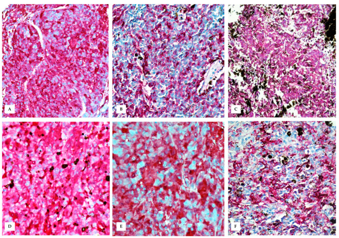
Figure 6.
Immunohistochemistry performed on skin tumour (A–C) and testis (D–F). (A) Immunohistochemical evaluation shows an intense red staining for HMB-45 (nuclear Mayer’s hemalum counterstain, 20×); (B) Melan-A (nuclear Mayer’s hemalum counterstain, 20×); (C) S-100 (nuclear Mayer’s hemalum counterstain, 20×); (D) Intense red staining for HMB-45 in testicular metastasis (nuclear Mayer’s hemalum counterstain, 40×); (E) Melan-A (nuclear Mayer’s hemalum counterstain, 40×); and (F) S-100 (nuclear Mayer’s hemalum counterstain, 40×).

