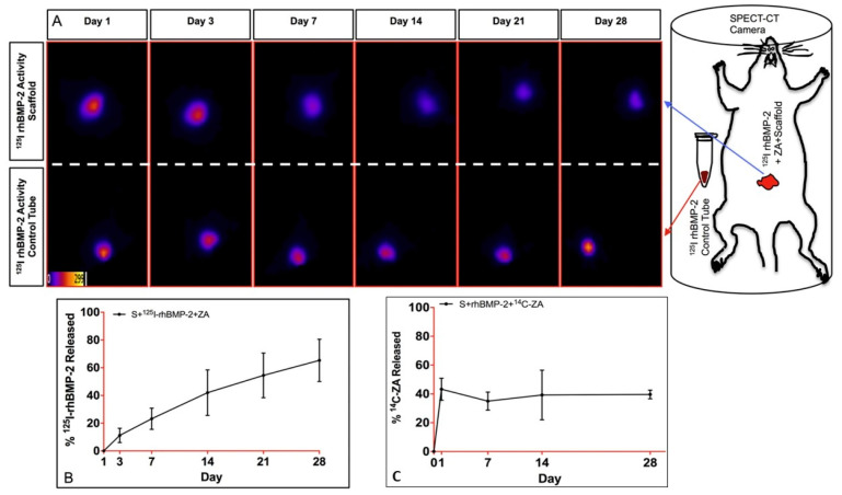
Figure 3.
In vivo 125I–rhBMP-2 release from the scaffolds in the abdominal muscle pouch model during 4-weeks via SPECT (single photon emission computed tomography)imaging. (A) SPECT signal from the scaffold containing 125I–rhBMP-2 + ZA in the abdominal muscle pouch (top (A)) and control tube containing known amount of 125I–rhBMP-2 placed outside the animal almost parallel to the implant (bottom (A)) at different time points. (B,C) % release kinetics of 125I–rhBMP-2 and 14C–ZA from the scaffold in the abdominal muscle pouch during the 4-week period. Adopted with permission from [68].

