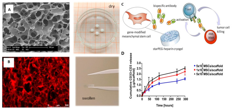
Figure 5.
(A) SEM image of PEG-heparin cryogel (left) and photograph of the cryogel scaffold in the dry state (right). (B) Confocal laser scanning microscopy image (left) and photograph of cryogel scaffold after swelling in PBS (right). (C) Illustration of the cryogel-housed scBsAb-releasing MSC system. (D) Release profile of bsAb CD33-CD3 from modified MSCs [87].

