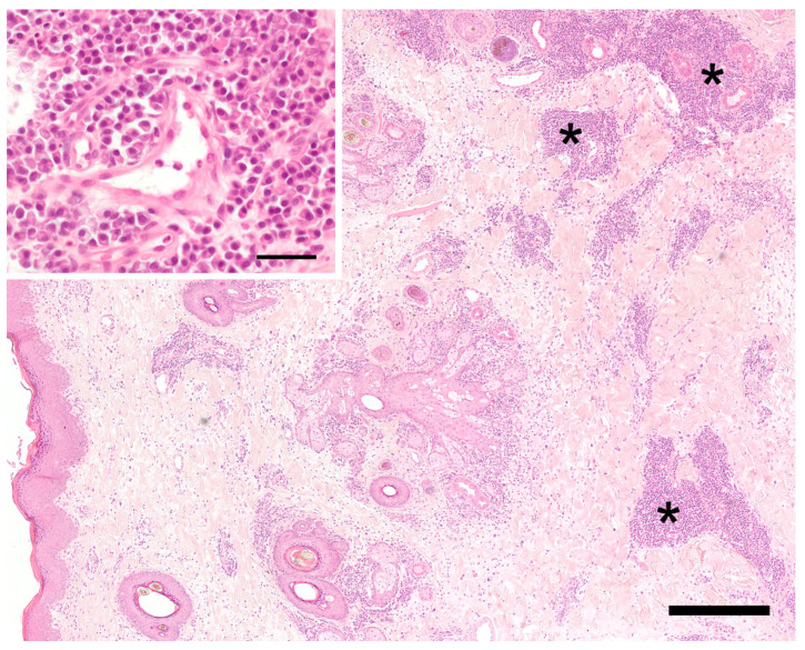
Figure 2.
Histology of the haired skin of the lip. The superficial and mid-dermis on the left show a moderate diffuse edema and dilated vascular and lymphatic vessels. On the right side, in the deep dermis and subcutis, there are perivascular and periadnexal, mainly plasmacellular inflammatory infiltrates (asterisks). Hematoxylin-eosin. Bar = 400 µm. Inset: perivascular plasmacells. Hematoxylin-eosin. Bar = 40 µm.

