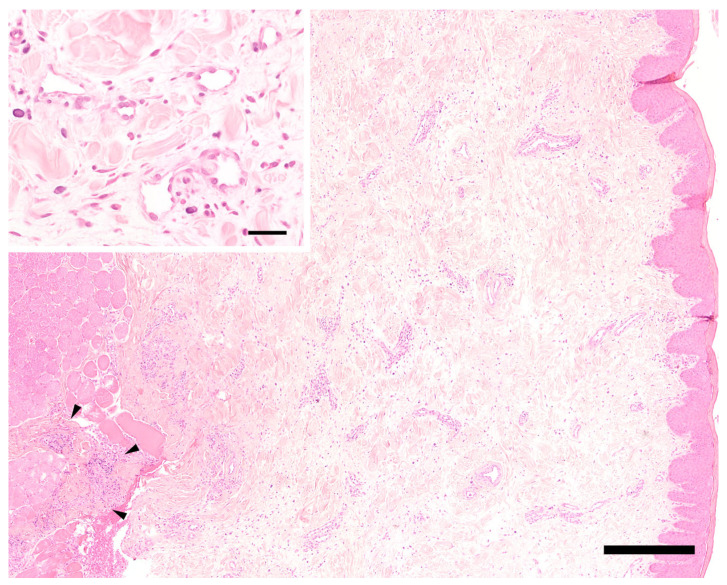
Figure 3.
Histology of the mucosa of the lip. Moderate diffuse edema is visible in the center and on the right side corresponding to submucosa and mucosa. Only few mixed perivascular inflammatory infiltrates are present, extending focally into the striated muscle on the left side (arrowheads). Hematoxylin-eosin. Bar = 400 µm. Inset: small caliber venous and/or lymphatic vessels are lined by plump endothelial cells. The vessel walls are otherwise unchanged. Note extensive clear spaces between tissue structures (extracellular edema). Hematoxylin-eosin. Bar = 40 µm.

