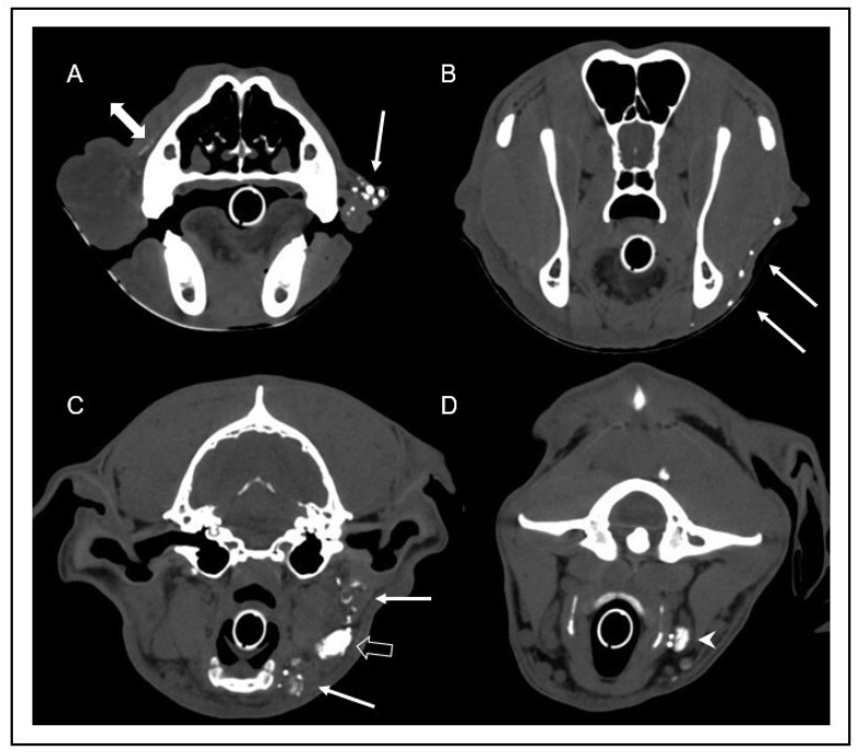
Figure 5.
Transverse computed tomographic images after indirect lymphography with subcutaneous injection of contrast medium into the rostral aspect of the bilateral lips one minute after injection. Images were obtained at the level of the rostral nasal cavity (A), frontal sinus (B), tympanic bulla (C), and atlas (D). Multiple well enhanced lymphatic vessels (arrows) can be identified in the left lip (A), continuing caudoventrally (B), and draining into the left mandibular lymph nodes (open arrow in image (C)) and the left medial retropharyngeal lymph node (arrowhead in image (D)). Note the scant volume of contrast media extending along the facial planes (double-headed arrow in image (A)). On the right side, lymphatic vessels could not be identified, and there was no contrast enhancement of the right mandibular and medial retropharyngeal lymph nodes.

